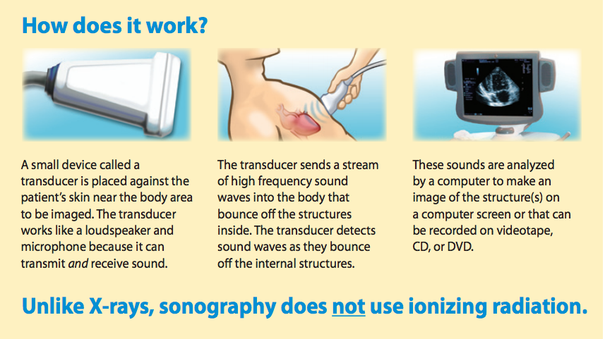
Diagnostic sonography (ultrasonography) is an ultrasound-based diagnostic imaging technique used for visualizing internal body structures including tendons, muscles, joints, vessels and internal organs for possible pathology or lesions.
It is a diagnostic medical procedure that uses high frequency sound waves to produce dynamic visual images of organs, tissues, or blood flow inside the body. This type of procedure is called a sonogram.
Types of Sonography
Abdomen: evaluation of all the soft tissues, blood vessels and organs of the abdomen
Breast: frequently used to evaluate breast lumps and other abnormalities that are found with screening or mammography
Obsterics/Gynecology: evaluation of the developing fetus and the female reproductive system
Cardiac: evaluation of the anatomy and function of the heart and related blood vessels
Vascular: evaluation of the blood flow through blood vessels
Neurosonology: evaluation of the brain and spinal cord in infants
Sonography is increasingly being used in the detection and treatment of heart disease, heart attack, and vascular disease that can lead to stroke. It is also used to guide needles for tissue biopsies taken for testing under a microscope. The professionals who perform these procedures are known as sonographers.